
So you just had your pet in to have their exam done with the veterinarian and were told they have dental disease and need to have their teeth cleaned. What does that mean exactly? Why do they need to be under a general anesthesia for that? You don’t see any wiggling teeth and they are eating fine, why is it they may need to have teeth pulled? Here is a more in depth look at what it means to have your pet’s’ teeth cleaned.
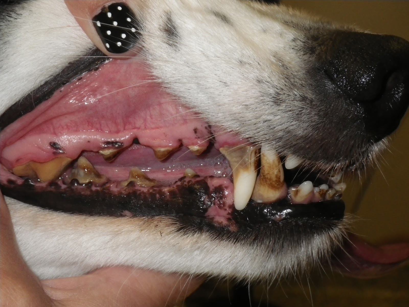
This dog has significant tartar buildup on his teeth as well as redness along the gum lines which is called gingivitis. He is in need of a dental cleaning.
As with any procedure that requires anesthesia, some preliminary bloodwork is always recommended. The purpose of bloodwork is to check the health of the pet’s’ internal organs that are responsible for filtering out the anesthesia. The results may change the doctor’s mind about what kind of anesthetic or pain medication to use. Even young pets can have internal organ compromise that can cause difficulty with anesthesia so it is recommended at any age.
Intravenous fluids are also highly recommended the day you pet comes in for the dental cleaning. Fluids will help keep blood pressure up. This is especially important if tooth extractions will be needed during the dental as they can take time so therefore your pet is under anesthesia longer. This helps wake your pet up faster as well as help flush out the internal organs of the anesthetic. It also gives the doctor direct access to the vein if any medications need to be given quickly during the procedure.

Our infusion pumps ensure your pet is getting the correct rate of fluids while they are in our hospital.
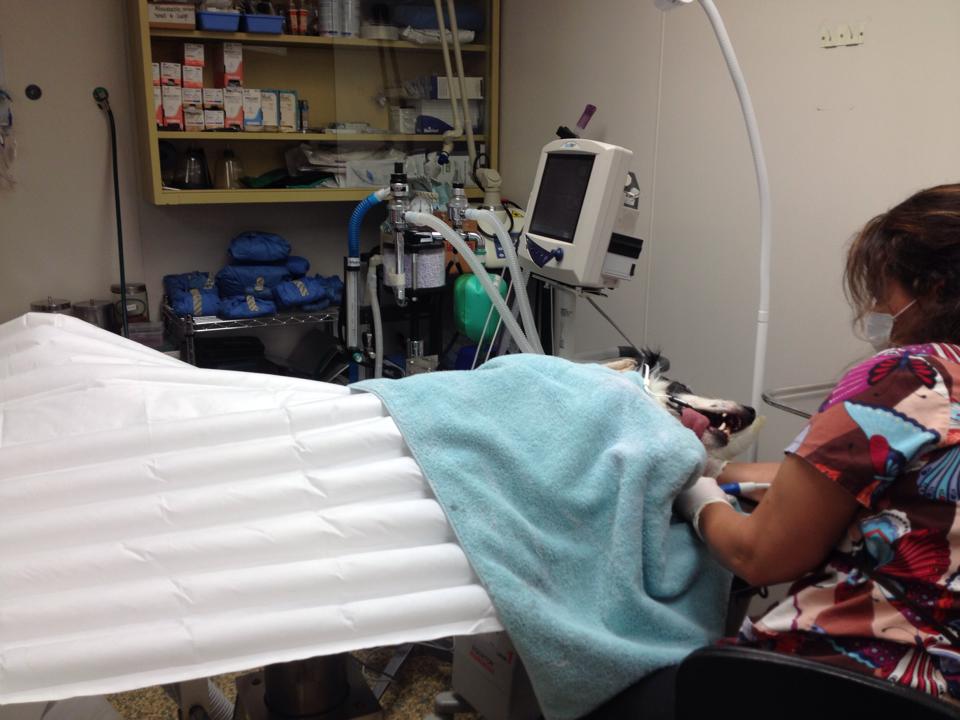
Once the doctor has determined the safest anesthesia for your pet depending on their bloodwork it is time for the dental to begin. A short acting injectable anesthetic is given that will allow the doctor to put into place an endotracheal tube to be able to maintain the pet on an anesthesia machine. This lets them monitor how much inhalant anesthesia they are receiving. This also allows the doctor to keep water out of the pets airways while the dental is being performed.
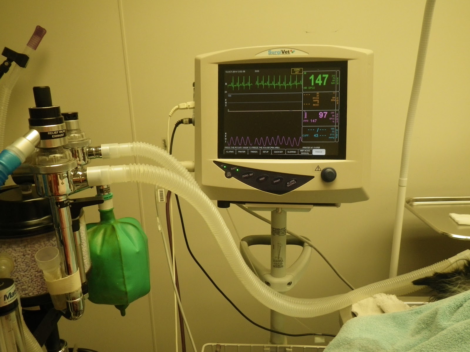
The pet also has their vitals monitored on our EKG monitor. This allows the doctor and technician to monitor heart rate, oxygen level, blood pressure and temperature throughout the procedure. We also have a hot air warmer that helps to maintain the temperature of the pet.
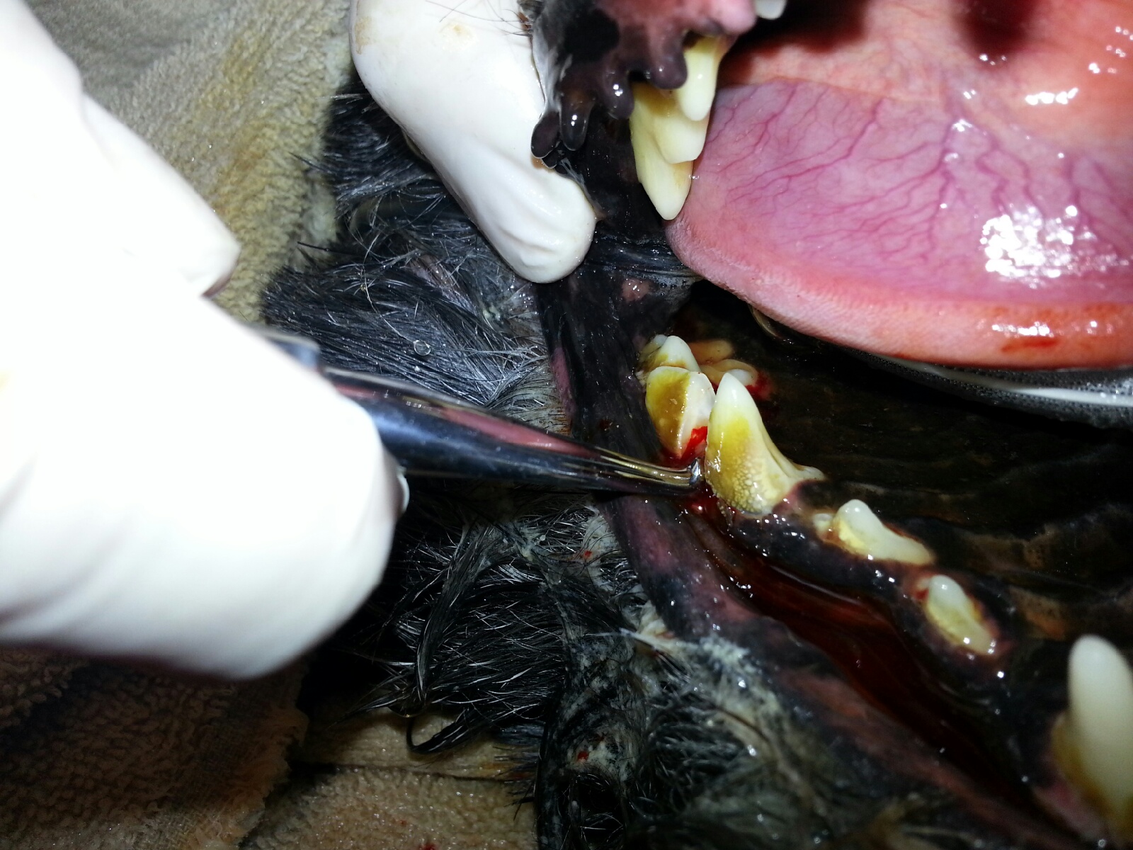
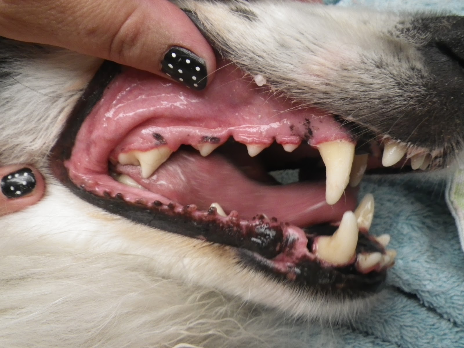
Once the pet is stable under anesthesia the doctor will check each tooth with a dental probe. What this means is, a very small marked probe is placed between the tooth and the gum. If the tooth is healthy, the probe should not go down between the tooth and gum very far, if at all. If a tooth is infected – or abscessed – this probe will fall further down in between the tooth and gum which means, this allows infection down to the root of the tooth and the tooth is diseased and needs to be extracted. This is not something that can always been visualized with them awake. Therefore, once they are asleep and the teeth are probed, there may be unexpected extractions needed. If the infected teeth are not removed, they just leave a constant source for pain and infection in the pet’s mouth and there is a very good chance that tooth will abscess at some point causing extreme pain and swelling in the mouth.
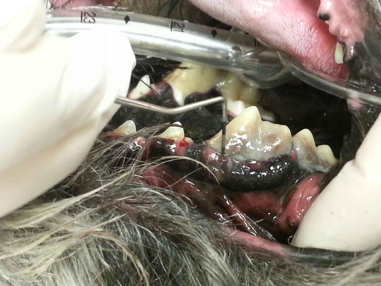

This shows an example of how when looking at the tooth, infection is seen but until they are asleep the tooth cannot be fully evaluated. When this particular tooth was checked once asleep, then dental probe can go fully through the tooth meaning the root is exposed. This is painful and the pet is constantly swallowing all that infection which can lead to heart, liver and kidney disease.
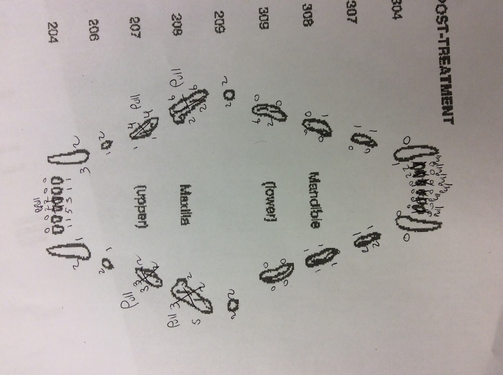
This is an example of a dental chart that is filled out for every dental. On it is written the pocket depth for each tooth, and any extractions or missing teeth. Once every tooth has been probed and charted on our dental chart, we then take full mouth radiographs to determine the health of the tooth roots.
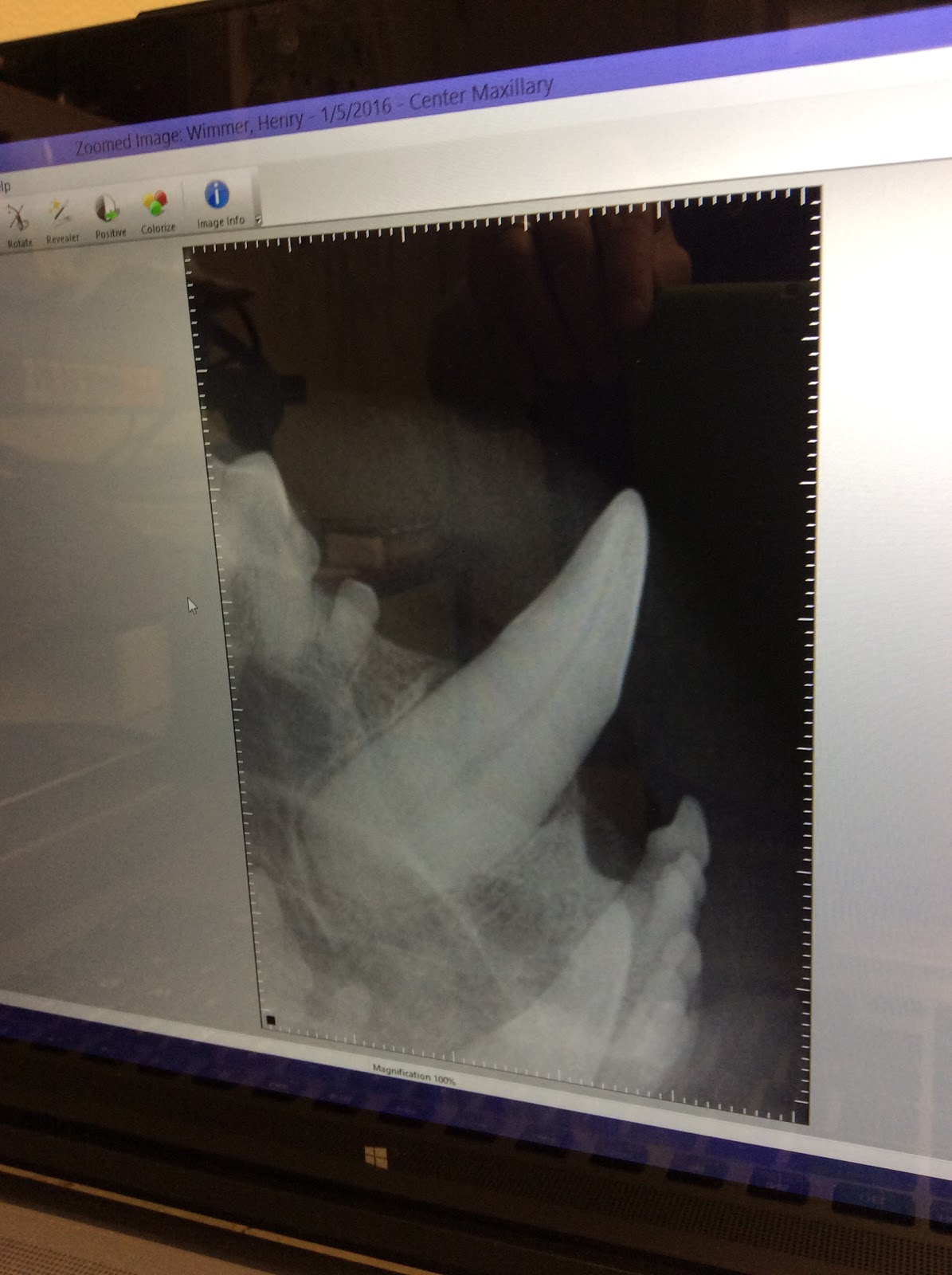
This is a radiograph of the large canine tooth. The entire root is healthy and in tact. No extraction is needed for this tooth.
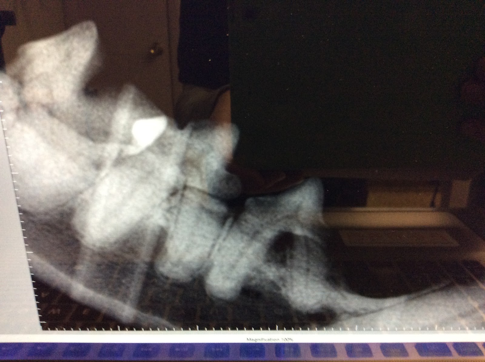
In this radiograph, the tooth on the right is unhealthy. The gum is receding away from the tooth leaving a space there for food and tartar to build up under the gum line as seen by the pocket or air between the two tooth roots. This tooth does need to be extracted.
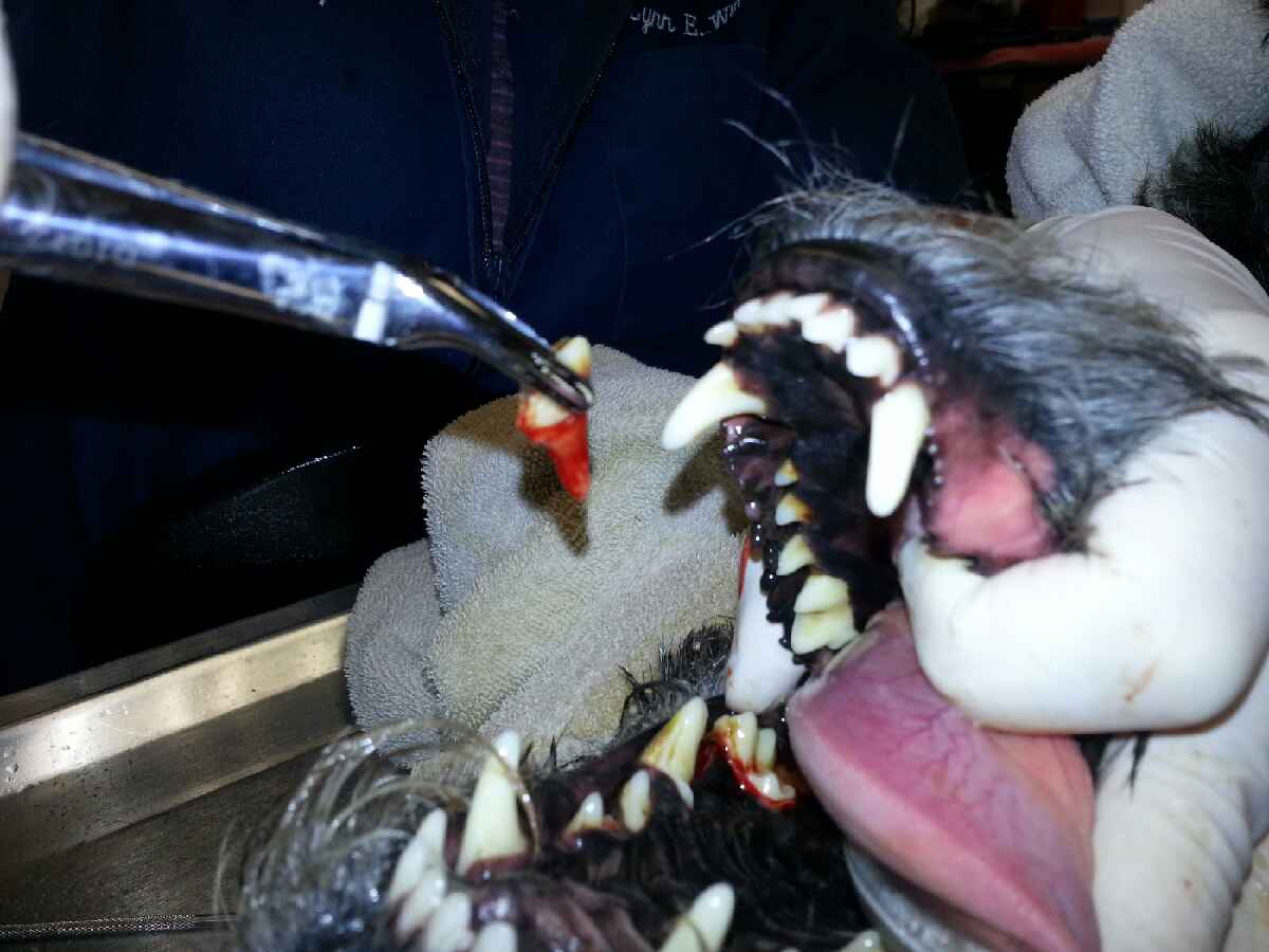
Once the probing and preliminary radiographs are done it is time for the doctor to perform any necessary extractions. Our dental machine has a high speed drill to assist the doctor with extractions. They will first section the tooth with the drill. This will allow the doctor to have better access to each root individually and ensure they remove the entire tooth root. Extractions can take time depending on which tooth is needed to be extracted and how abscessed they are.

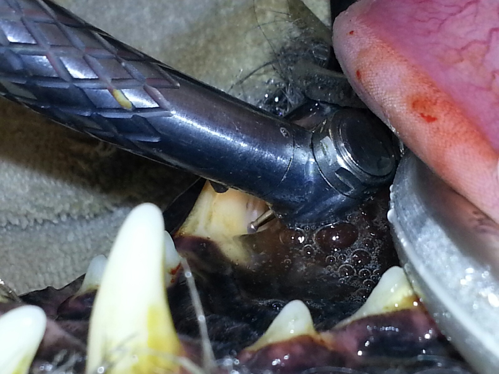
Once the tooth is sectioned the doctor will use elevators to loosen the ligaments that hold the tooth in place to be able to remove the tooth entirely.

Once all extractions are done, radiographs are taken again to ensure there are no roots or fragments left under the gum line that could cause problems later. In this radiograph the middle 2 teeth were removed and there are no remaining roots or fragments.
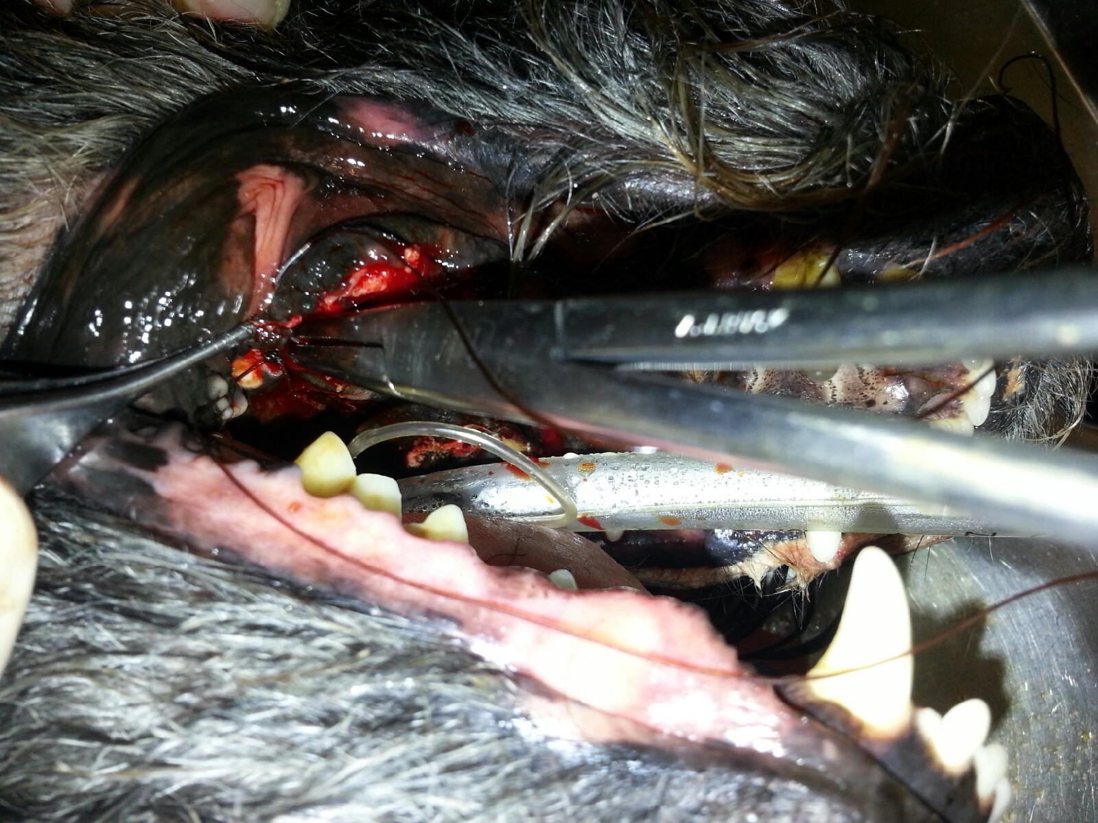
Now that the tooth is removed, the doctor sutures the gum closed so that food and particles cannot get into the socket where the tooth was and cause any pain or infection.
After the doctor is finished with any necessary extractions it is time for the teeth to be scaled and polished. Our dental machine has an ultrasonic scaler to break off the tartar on the teeth and get them as clean as possible. There may be tooth staining that cannot be removed, but the tartar buildup on every tooth, inside and out, is removed. When scaling teeth, tiny microscopic grooves are left on the tooth which can cause food and debris to adhere to. This is why polishing is so important. Polishing smoothes out the tooth so that the food and debris cannot adhere as quickly to the teeth.

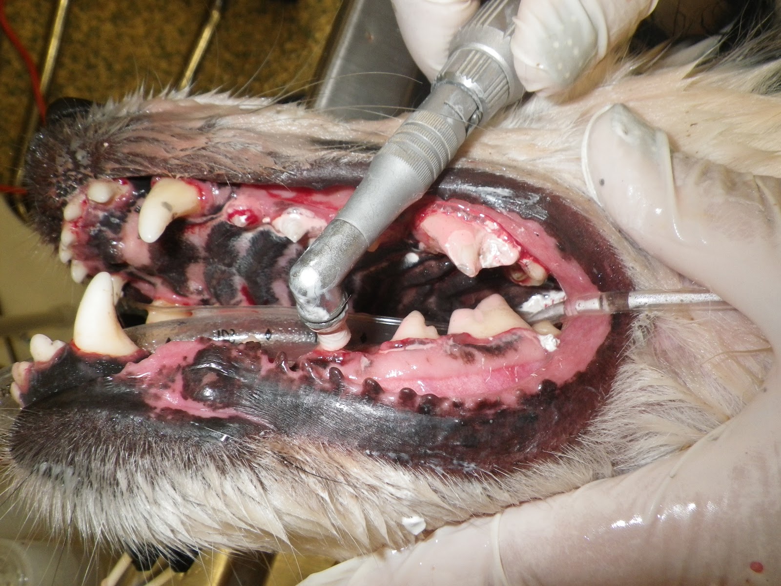
Scaling the teeth Polishing the teeth
And here is the finishing product. A beautiful set of teeth!